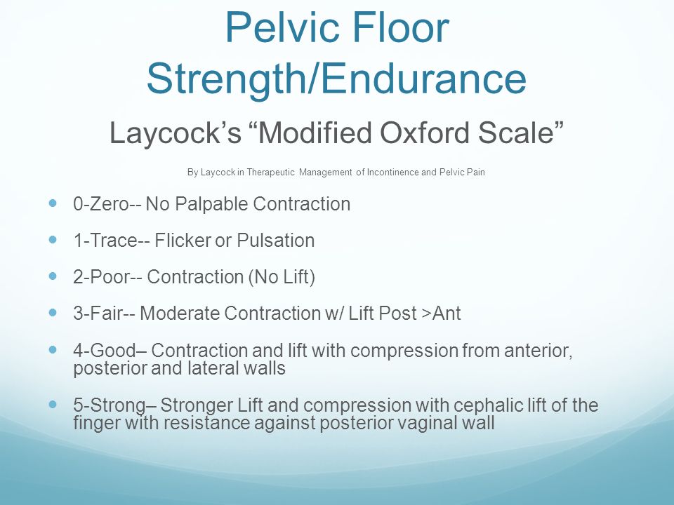
Pelvic Floor Strength Scale

Kegel8 Squeeze Scale From The Kegel8 Trainer Kegel8

Table 1 From Measurement Of Pelvic Floor Muscular Strength With The Colpexin Pull Test A Comparative Study Semantic Scholar

Pelvic Floor Exercises During Pregnancy Pregnancy Yoga Newmarket

Pelvic Floor Muscle Strength In Prostate Cancer Patients Treated With Download Table
Physiotherapy First For Pelvic Floor Dysfunction Urology News

Visionwowgroup
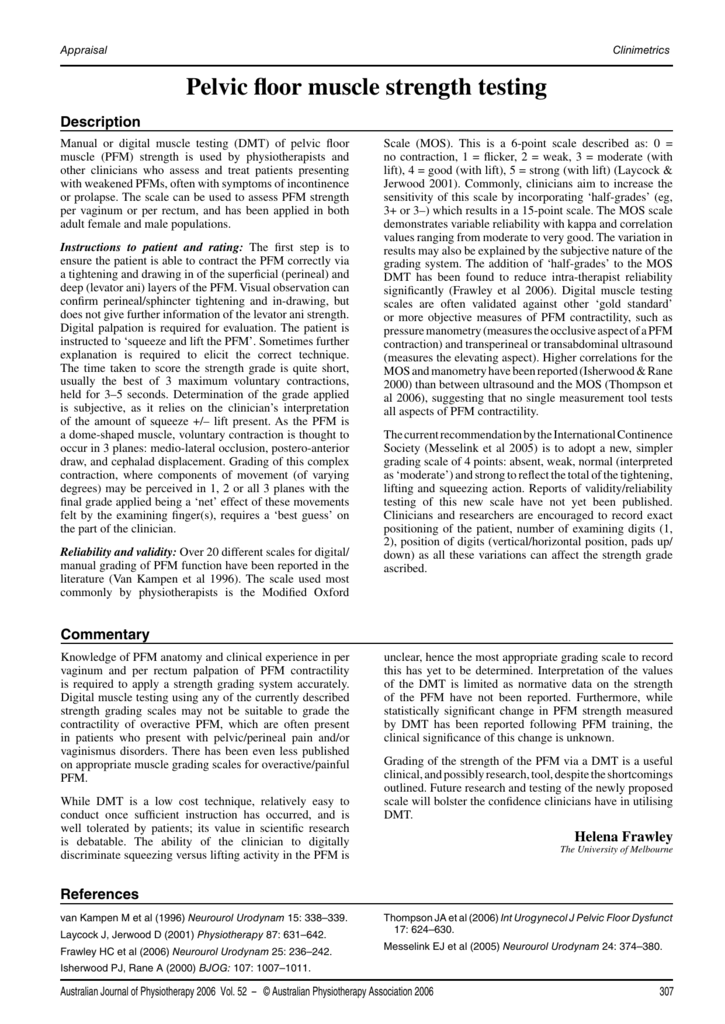
To develop a digital technique to assess pelvic floor muscles pfm.
Pelvic floor strength scale.
Http Www Hkag Org Conference Incontinence 20symposium Notes Hkcs Pdf

Table 2 From Oxford Grading Scale Vs Manometer For Assessment Of Pelvic Floor Strength In Nulliparous Sports Students Semantic Scholar

Protocol For Pelvic Floor Muscle Training Download Table

Women Understanding Preventing And Managing Pelvic Floor Dysfunction Kathleen Zonarich Pt Ppt Video Online Download

Therapy For Management Of Childbirth Perineal Tears And Post Partum Pain Page 3

Using The Brink Score To Predict Postpartum Anal Incontinence Semantic Scholar
Pdf Normal Reference Values Of Strength In Pelvic Floor Muscle Of Women A Descriptive And Inferential Study
Introduction To Women S Health Physical Therapy Tara Sullivan Pt Dpt Prpc Ppt Download
Pdf Evaluation Of Female Pelvic Floor Muscle Function And Strength

Digital Assessment Of The Pelvic Floor Muscles A Neglected Technique
Pelvic Floor Muscle Strength Testing

A Urogynecology Review For Up College Of Medicine Interns
Pelvic Floor Muscles Pelvic Floor Muscles Grading

Pdf Inter Rater Reliability Study Of The Modified Oxford Grading Scale And The Peritron Manometer Semantic Scholar
Pelvic Floor Muscle Function And Strength Physiopedia

Full Text Questionnaires To Evaluate Pelvic Floor Dysfunction In The Postpartum Ijwh

Pdf The Effects Of Pilates Method On Pelvic Floor Muscle Strength In Patients With Post Prostatectomy Urinary Incontinence A Randomized Clinical Trial

Pdf Pelvic Floor Muscle Training During Pregnancy And After Delivery
Https Encrypted Tbn0 Gstatic Com Images Q Tbn 3aand9gcqzwbbx59 Lrwm7zdkpzglcjywzby6bgjl Vpulvzvahcx4gthr Usqp Cau

Effects Of Multidimensional Pelvic Floor Muscle Training In Healthy Young Women Semantic Scholar

Pelvic Floor And Abdominal Muscle Cocontraction In Women With And Without Pelvic Floor Dysfunction A Systematic Review And Meta Analysis
Https Www Ics Org Workshops Handoutfiles 000893 Pdf

Causes Of Pelvic Floor Muscle Dysfunction Pfmd Download Scientific Diagram
Physiotherapy Assessment For Female Urinary Incontinence Springerlink
Source : pinterest.com
